Fluorescence imaging is a versatile and sensitive method which allows non-invasive investigations of both fossils and minerals. All minerals have unique optical properties and by capturing multiple fluorescence images with different light and filter combinations, similar and dissimilar surface layers that are not easy to spot by visual examination are quickly identified. We will show how advances in fluorescence technology can be used for characterization of fossils.
Introduction
Physical evidence of life’s rich history on Earth is preserved in minerals and rocks as fossils. Fossils come in all shapes and forms, from bones to imprints of organic material. Careful examination of the fossil and its site of discovery helps determine the age and environment of the fossil. Numerous scientific methods are used for analysis of the fossil, including:
- Light microscopy to view fine details
- Electron microscopy to view details at high resolution
- Vibrational spectroscopy to map chemical compounds on the surface
- X-ray and CT scanning to investigate internal structures
- Isotope analysis to study diet and habitat
- Radiometric dating
- DNA analysis to study evolutionary relationships
- Fluorescence imaging
Recent advances in fluorescence imaging have introduced new light sources and emission filters, from UV to near-infrared (NIR), which opens up new avenues of exploration for a wide range of sample types. We show how two imaging systems, Amersham™ ImageQuant™ 800 imaging system and Amersham™ Typhoon™ laser scanner, can be used for the investigation of fossil samples (Fig 1). Both instruments are well suited for fluorescence analysis of a wide variety of specimens.
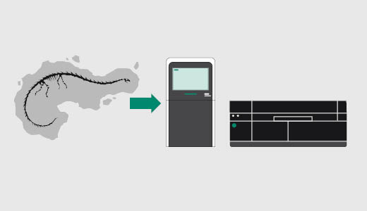
Fig 1. We used the Amersham™ ImageQuant™ 800 imaging system and Amersham™ Typhoon™ scanner to image fossils, revealing new information about the specimens.
Due to the fragility of fossils some preservation is usually needed. Cleaning, gluing, and sometimes stabilizing with a protective coating aid with preservation but may complicate the surface analysis. Also, in some cases fossils have been artificially improved and faked. For those cases, fluorescence imaging is a useful method to help distinguish fossils from other man-made materials (1).
Fluorescence imaging is a quickly evolving field, with many different options available. The Amersham™ ImageQuant™ 800 imaging system allows a great number of different combinations of excitation LED light and band-pass emission filters. The six built in excitation LEDs cover a spectral range from UV (365 nm) to near infrared (775 nm). The working area (16 × 22 cm) allows for small to medium sized fossils to be investigated. Furthermore, the efficient light collection allows for a numerical aperture with a large depth of view. Thus, non-flat objects are well suited for investigations with the Amersham™ ImageQuant™ 800 imaging system. While, for flat samples < 2 cm in height, the Amersham™ Typhoon™ laser scanner can be used. The Amersham™ Typhoon™ laser scanner also has a large scan area of 40 × 46 cm, and is able to achieve high resolution with pixel sizes below 100 micrometers (Table 1).
Table 1. Comparison of imagers used to image fossils
| Amersham™ ImageQuant™ 800 imaging system | Amersham™ Typhoon™ laser scanner | |
| Scan area | 16 × 22 cm | 40 × 46 cm |
| Sample requirements | • Light is collected from above the sample • Non-flat samples can be imaged with fluorescence depth of view of approximately 2 cm |
• Flat samples facing the scanner glass plate • Max physical sample height: 2 cm |
| Light sources | LED: 365, 460, 535, 635, 660, 775 nm + white light sources | Lasers: 488, 532, 635, 685, 785 nm |
| Detector | 8.3 megapixel cooled CCD | High dynamic range PMT |
| Output files | 16-bit analysis files, plus color images | 16-bit analysis files, no white light color images |
Results and discussion
Overview of investigated fossils
Fossil specimens were loaned courtesy of the Museum of Evolution, Uppsala University, Sweden. The fossil selection was chosen to represent a wide span of fossil types from different eras. The youngest fossil was the insect Dipteran encased in amber from the Eocene epoch 34 to 56 million years ago, and the oldest was from the Cambrian era. The selection of fossils we examined is listed in Table 2.
Table 2. The fossils examined in this study had been collected in many locations and originate from different time periods (All fossil specimens were loaned courtesy of the Museum of Evolution, Uppsala University, Sweden.)
| Specimen | Description | Figure |
| Dipteran | Insect encased in amber from the Baltic region | 3 |
| Stellostomites | Enigmatic Cambrian fossil from Chengjiang, China | 4 |
| Asaphus | Ordovician trilobite from Östergötland, Sweden | 5 |
| Alethopteris | Carboniferous fern from Germany | 6 |
| Phalarodon | Triassic ichtyosaurian marine reptile from Svalbard | 7 |
| Pachylpleurosaurus | Triassic sauropterygian marine reptile from Italy | 8 |
| Keichousaurus | Triassic sauropterygian marine reptile from China | 9 |
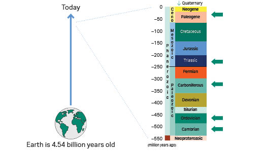
Fig 2. Timeline and different fossils examined in this study indicated with green arrows. The current geologic eon covers a time span of 541 million years, and it began with the Cambrian Period when animals first developed hard shells which could be preserved in the fossil record.
Near infrared fluorescence (NIR) reveals new details
The fact that many materials do not absorb NIR light makes NIR fluorescence less sensitive to background. This can be used to resolve features at greater depth in the sample and is used, for example, in biomedicine and tissue examinations. This is also a useful feature for examining fossils.
The first fossil we examined was Dipteran encased in amber from the Baltic region (Fig 3). We observed no signal of the insect with visible light excitation but with NIR excitation it was easy to detect auto-fluorescence from the insect. The exact shape of the insect was distorted in the image due to the non-flat surface of this amber. This shows that NIR fluorescence is a promising method to study objects in ambers, which are semi-transparent.
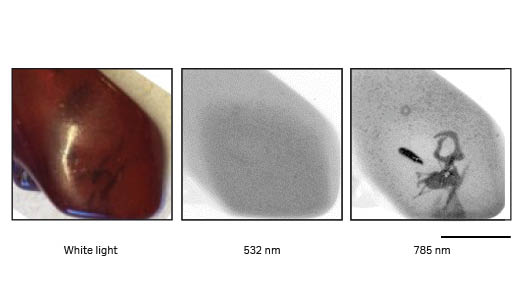
Fig 3. Images of polished amber with insect (Dipteran) from the Baltic region. The insect inclusion was not detectable with visible fluorescence excitation (532 nm; 570BP20) but clearly detected with NIR excitation (785 nm; 825BP30) using the Amersham™ Typhoon™ laser scanner. The curved surface of the amber lead to some image aberration. The black scale-bar is 1 cm. The contrast of the grayscale images ranges from white (low intensity) to black (high intensity).
Next, we investigated a Stellostomites fossil from the Cambrian time period. This was the oldest fossil we studied, and it exhibited strong auto-fluorescence in the NIR region of the spectrum when imaged using the Amersham™ ImageQuant™ 800 imaging system (Fig 4).
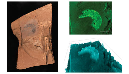
Fig 4. White light (left) and fluorescence images (upper right) of a Stellostomite fossil from Chengjiang, China (Cambrian). Amersham™ ImageQuant™ 800 imaging system was used to image the fossil at NIR excitation (775 nm). The scale bar is 1 cm. A 3D view (lower right) can be useful to visually assess the signal distribution across a surface.
We also investigated an Asaphus trilobite to examine a specimen with a non-flat topology. We found evidence of differential fluorescence emission at the head of the trilobite, which suggests there was some difference in chemical composition (Fig 5).
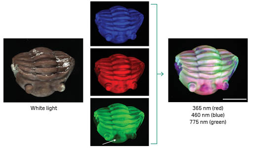
Fig 5. White light and fluorescence multi-color images of a trilobite (Expansus-layer, Ordovician), Närke, Sweden. Different parts and layers of the fossil were examined using different light and filter combinations of the Amersham™ ImageQuant™ 800 imaging system. The NIR excited fluorescence (775 nm) differ in some part of the region between the eyes of the trilobite, indicated by the arrow. The white scale bar is 1 cm.
Very large Stokes shifts measured with UV illumination and NIR band-pass filter
Long periods in extreme conditions, pressed between rocks, often transform organic material to thin carbon films. These fossils are well suited for non-contact fluorescence imaging. We examined a fern fossil from the Carboniferous era (Fig 6). We observed a fluorescence emission with a very large Stokes shift. The fern fossil was illuminated with 365 nm UV LED, and emitted fluorescence around 840 nm was detected with a band-pass filter, which corresponds to a Stokes shift of almost 500 nm. Further investigations are needed to understand what type of carbon the fossil consists of, which is often done with vibrational spectroscopy.
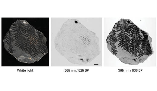
Fig 6. White light and fluorescence images of Alethoperis carbon film fossil (Germany). Amersham™ ImageQuant™ 800 imaging system with UV excitation at 365 nm and collection at 525 nm (middle image) and 836 nm (right image) using a band-pass filter was used to show how different layers of a fossil can be examined with different light and filter combinations. The contrast of the grayscale images ranges from white at the weakest intensity to black. The scale bar is 1 cm.
Easy area comparisons
One of the most useful features of fluorescence imaging is the ease at which different regions can be compared. Most materials emit autofluorescence and similar materials emit light with similar intensity. Especially when comparing several images in a color-overlay image it is easy to identify regions of similar chemical composition. One example is a Phalaradon fossil found at Svalbard (Fig 7). The green and purple areas of the fossil color overlay indicate differences in chemical composition. There is also an area in the upper right corner which could come from a different fossil.
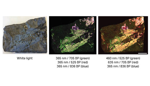
Fig 7. White light and fluorescence overlay images, using Amersham™ ImageQuant™ 800 imaging system, of a partial skull of Phalarodon Nordeskioldii, Middle Triassic (Ladinian), “Upper Saurian niveau,” Botneheia Formation, Spitsbergen. Different excitation light and band-pass filter combinations reveal layers of similar material in the fossil. For example, the teeth outline exhibits a strong fluorescence signal indicated with the purple color. The white scale bar is 20 mm.
Using the Amersham™ Typhoon™ laser scanner to illuminate finer details
The Amersham™ Typhoon™ laser scanner can be used to image fossils which are flat and not too thick (max 2 cm in height). The Amersham™ Typhoon™ laser scanner provides a higher resolution and a more specific excitation due to its optics design. Protective padding around the fossil area is recommended to safeguard the fossil and scanner. The well-preserved specimen in Figure 8 was scanned with a resolution of 10 µm per pixel. The wealth of high-resolution information of these images allows detailed investigations of the anatomy of this specimen at sub mm level.
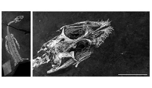
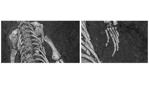
Fig 8. The fossil Pachylpeurosaurus staubi (Middle Triassic) from Besano Formation, Italy exhibits strong fluorescence signals in all fluorescence channels. The contrast of the grayscale images ranges from black (weak intensity) to white at the strongest. The images were captured with the Amersham™ Typhoon™ laser scanner using a 488 nm laser at 10 µm pixel size. The scale bar is 1 cm.
Finally, with increasing commercial interest in fossils, it is becoming more common with artificial fossil improvements and even fake fossils. It can be time-consuming to investigate a fossil in such detail to decide if this is the case. Fluorescence imaging allows for a quick screening of authenticity. In particular, expected autofluorescence levels based on investigations of similar fossils can be used as a first criteria. The level of detail and distribution of fluorescence intensity is also a useful quality indicator. An example of a fossil that requires further investigation is shown in Figure 9. It is potentially Triassic sauropterygian marine reptile fossil but it lacks fine details, compared to the fossil in Figure 8, and the auto-fluorescence was weak in all channels.
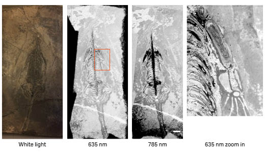
Fig 9. The fossil Keichousaurus hui exhibited weak and diffuse fluorescence signals using the Amersham™ Typhoon™ laser scanner. The contrast of the grayscale images ranges from white at the weakest intensity to black. The white scale bar is 1 cm.
Conclusion
We have shown that modern imaging systems, such as the Amersham™ ImageQuant™ 800 imaging system and Amersham™ Typhoon™ laser scanner, can be used for the characterization of fossils. Modern imagers offer a wide variety of options in terms of light sources and light collection filters.
- The autofluorescence of some material in the NIR region allows you to see deeper into materials, such as, with the amber and trilobite.
- In the study of carbon films, UV excitation and emission with very large Stokes shift can be used to study amorphous carbon phases.
- Fluorescence imaging also allows for quick comparisons of different surface layers.
- Finally, the Amersham™ Typhoon™ laser scanner allows for careful examination of fine details in well-preserved specimen.
References
- Selden PA, Olcott AN, Downen MR, Ren D, Shih C, Xiaodong C. The supposed giant spider Mongolarachne chaoyangensis, from the Cretaceous Yixian Formation of China, is a crayfish, Palaeoentomology. 2019;2(5):515-522.
CY36444-27Nov23-AN